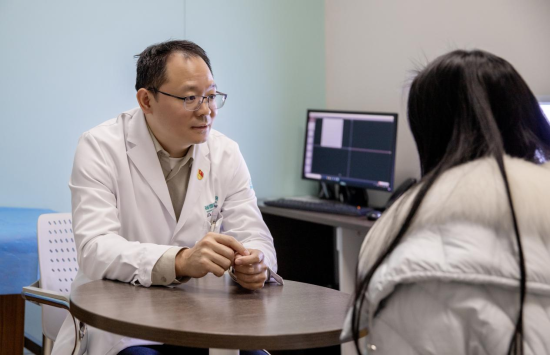
Dr. Gai Yanting discusses treatment options with a patient at Shanghai Donglei Brain Hospital.
Children & Teens
Hemorrhage
Complications
A Routine PE Class Turns into an Emergency
For 18-year-old Wen Hua (a pseudonym), it started like an ordinary school day. During a PE class, she suddenly felt a severe headache, followed by nausea and the urge to vomit. She rested, but the headache did not ease. Instead, it grew worse, and she began vomiting repeatedly.
At the hospital, imaging revealed the real cause: brain arteriovenous malformation (AVM) with rupture and bleeding. The diagnosis shocked her family. She was transferred to Shanghai Donglei Brain Hospital, where Dr. Gai Yanting, Director of Neurosurgery, led her care.
"This type of vascular malformation is particularly difficult to treat because it is located deep in the brain and within important functional areas. Open surgery carries high risk and a significant rate of complications."
What Is a Brain AVM?
A brain arteriovenous malformation (AVM) is a congenital abnormality of cerebral blood-vessel development. In a healthy brain, arteries deliver oxygen-rich blood through a network of tiny capillaries to veins. In an AVM, arteries connect directly to veins through a tangled nidus of abnormal vessels, bypassing the capillary bed.
Without capillaries to buffer pressure, the venous side is exposed to high arterial pressure. The nidus often contains enlarged, aneurysm-like veins. This abnormal hemodynamics makes AVMs prone to rupture and intracranial hemorrhage.
Key point: A brain AVM is usually present from birth. Most people do not know they have one until it bleeds.
Why Venous Embolization Was Chosen
Wen Hua's AVM sat in a deep, eloquent area of the brain. Conventional open resection would have carried high risk of neurological injury. After multidisciplinary discussion, Dr. Gai Yanting's team chose a transvenous embolization approach.
Embolization is a minimally invasive neurointerventional procedure. A microcatheter is threaded through the venous system into the AVM nidus, and embolic material is injected to block the abnormal shunts. For Wen Hua, this approach offered a better balance between treating the lesion and preserving brain function.
1. Minimally Invasive
No large scalp incision or skull opening. Access is gained through a small puncture in the groin or wrist, reducing surgical trauma.
2. Suitable for Deep or Eloquent Lesions
When an AVM is located near speech, motor, or visual pathways, catheter-based treatment can be safer than open surgery.
3. Faster Recovery
Patients typically spend less time in the hospital and return to daily activities sooner than after craniotomy.
Outcome: Back to Normal Life
Using their extensive experience in cerebrovascular disease, Dr. Gai Yanting's team successfully delivered embolic material into the nidus and completely blocked the abnormal blood flow. After the procedure, Wen Hua improved significantly and developed no complications. She was able to return to her studies and normal daily life.
When Treatment Is Recommended
Specialists individualize AVM treatment based on the patient's condition and the lesion's anatomy. Dr. Gai emphasizes that patients with a prior hemorrhage or high-risk features for rebleeding should generally receive active intervention, especially when symptoms are present.
⚠️ What Triggers an AVM Rupture?
Many factors can raise blood pressure or stress the abnormal vessels and trigger AVM bleeding. Common triggers include:
- Strenuous exercise or sudden physical exertion
- Emotional excitement or intense stress
- Physical fatigue and sleep deprivation
- Alcohol consumption
- High blood pressure or sudden spikes in pressure
- Straining, heavy lifting, or breath-holding
- Weather changes or sudden temperature shifts
However, some AVMs rupture without any obvious trigger, even while the person is at rest.
Red-Flag Symptoms: When to Seek Emergency Care
Because brain AVM is congenital, preventing it from forming is not possible. But recognizing the warning signs of rupture and acting fast can save lives. Dr. Gai urges anyone with the following symptoms to seek immediate medical evaluation:
- Sudden, severe headache — often described as the "worst headache of my life"
- Dizziness, nausea, or vomiting
- Seizure with no prior history
- Numbness, weakness, or paralysis on one side of the body
- Vision changes, slurred speech, or confusion
- Loss of consciousness
Early diagnosis through imaging allows doctors to classify the AVM, estimate rupture risk, and choose the safest treatment pathway.
How Brain AVM Is Diagnosed and Treated
Diagnostic workup usually begins with emergency brain imaging. Once the patient is stable, additional vascular imaging helps define the AVM's size, location, feeding arteries, and drainage pattern. Common tools include:
- CT scan — first-line emergency imaging to detect bleeding
- MRI — detailed visualization of the nidus and surrounding brain
- Cerebral angiography — the gold standard for mapping blood supply and drainage
Treatment options include:
- Open microsurgery — complete removal of the AVM
- Endovascular embolization — catheter-based occlusion
- radiosurgery — focused radiation to close the nidus over time
- Combination therapy — two or more modalities used together
The best approach depends on the patient's age, symptoms, AVM size and location, and risk of future bleeding.
Know the Signs, Act Fast
Wen Hua's case is a powerful reminder that a sudden severe headache in a young person is never "just a headache" until proven otherwise. At Shanghai Donglei Brain Hospital, timely diagnosis and a carefully chosen venous embolization gave her a second chance. As Dr. Gai puts it, "When it comes to AVM, the first bleed is a warning. The goal is to prevent the next one."
Concerned About a Sudden Severe Headache or AVM?
Shanghai Donglei Brain Hospital offers 24/7 emergency neurovascular evaluation, advanced brain imaging, and minimally invasive treatment options.
Request a Consultation