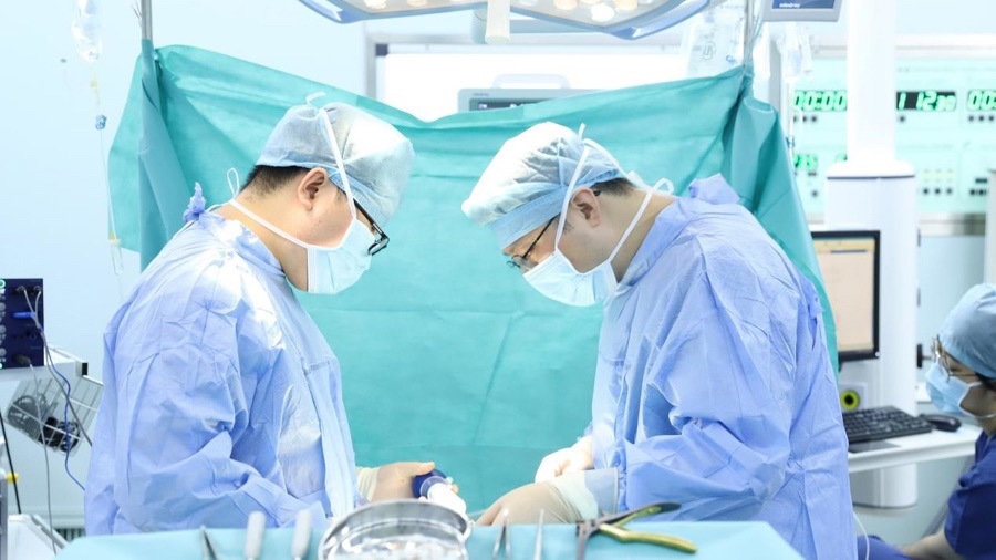
Dr. Wang Wei and team during skull-base tumor surgery at Shanghai Donglei Brain Hospital. (Image: Shanghai Observer)
In This Article
A Blurry World: When Vision Loss Hides a Brain Tumor
For nearly two years, 55-year-old Mr. Zhou (a pseudonym) noticed his eyesight growing steadily worse. At first, he dismissed it as a normal part of aging. "I thought it was just presbyopia," he recalled. Eventually, his left eye could barely perceive light and shadow, and the world on his left side seemed to disappear.
It was only after a cranial MRI that the real culprit was found: not a problem with the eye itself, but a giant skull-base meningioma measuring approximately 8×7 cm, pressing on the optic pathways and displacing cranial nerves.
40% of Sellar Meningiomas First Visit an Eye Clinic
Meningioma is one of the most common primary intracranial tumors in adults, accounting for about 40% of all primary intracranial tumors. "Meningiomas in the sellar and anterior skull-base region often compress the optic chiasm or optic nerve first," explained Dr. Wang Wei, Director of Neurosurgery at Shanghai Donglei Brain Hospital. "The earliest symptom is progressive, often one-sided vision loss."
"Approximately 40% of sellar meningioma patients are first seen in an ophthalmology clinic. They are often misdiagnosed with glaucoma, optic neuritis, or simple refractive problems. By the time an intracranial tumor is discovered, the visual damage is usually already severe."
Mr. Zhou's vision loss was not generalized blurring; it was a gradual shrinking of the left visual field. He began to miss doorframes on his left side. Dr. Wang explained that visual-field defects caused by tumor compression are typically unilateral and progressive — like a curtain slowly closing from one side.
👀 Why Meningiomas Hide in Eye Clinics
- Early target: Sellar and anterior skull-base meningiomas compress the optic chiasm or optic nerve first.
- First symptom: Progressive, often one-sided vision loss rather than eye pain.
- Common misdiagnoses: Glaucoma, optic neuritis, cataract, or routine refractive error.
- High stakes: Delayed diagnosis can lead to irreversible optic-nerve damage.
Starve First, Remove Second: Combined Embolization and Surgery
Mr. Zhou's tumor filled the sellar region and extended into the left frontal-temporal area. It had tightly encased several major intracranial "lifeline vessels" — the internal carotid artery, anterior cerebral artery, middle cerebral artery, and basilar artery — and displaced the optic chiasm and oculomotor nerve. The tumor was not simply sitting in the brain; it was embedded in a dense jungle of nerves and blood vessels at the skull base.
Dr. Wang Wei's team designed a "starve first, remove second" combined strategy:
The two-step plan:
- Step 1 — Preoperative embolization: Embolic material is injected into the tumor's feeding arteries, dramatically reducing its blood supply.
- Step 2 — Open surgical resection: Once blood flow to the tumor is markedly reduced, the tumor is carefully separated from the surrounding arteries and nerves and maximally safely removed.
- Result: Reduced intraoperative bleeding, shorter operative time, and a higher chance of total resection.
"For large skull-base meningiomas over 6 cm in diameter with rich blood supply, preoperative embolization can significantly reduce intraoperative blood loss, shorten surgical time, and improve the gross total resection rate," said Dr. Wang.
Will Vision Return? It Depends on the Nerve
After surgery, Mr. Zhou recovered better than expected. His left eye could already recognize faces.
"Whether vision recovers depends on whether the optic nerve has been compressed or destroyed. Mr. Zhou was fortunate — his nerve was compressed, not destroyed."
If the tumor merely compresses nerve fibers, removing the pressure may allow partial or even complete functional recovery. However, if compression has persisted too long, causing nerve atrophy or demyelination, the room for recovery becomes very limited.
Patient Age
Meningioma Size
First Seen in Eye Clinics
Resection Strategy
Warning Signs That Should Prompt Brain Imaging
Dr. Wang Wei reminds patients that both meningioma and arteriosclerotic stenosis develop over time. "A 55-year-old patient's vascular and tumor-related disease may have roots stretching back more than a decade."
⚠️ Symptoms Often Dismissed as Aging or Fatigue
- Unexplained vision decline or visual-field defects, especially on one side.
- Morning headaches that worsen with straining or coughing.
- Unexplained decrease in sense of smell.
- Unilateral hearing loss or persistent tinnitus.
- Personality changes, slowed responses, or obvious memory decline.
For middle-aged and older adults with hypertension, diabetes, or smoking history, a routine physical that only includes a neck ultrasound is not enough for brain health. A more comprehensive cerebrovascular and neuroimaging evaluation — such as MRI/MRA or CT/CTA — is essential. Early detection and early intervention are the keys to preventing avoidable vision loss or neurological damage.
🏆 Seeing the True Cause Behind the Symptoms
Mr. Zhou's case underscores a critical message: vision loss is not always an eye problem. A giant skull-base meningioma can present with symptoms that look like ordinary aging. Through accurate diagnosis, preoperative embolization, and meticulous skull-base surgery, the team at Shanghai Donglei Brain Hospital removed the tumor and gave his vision a chance to recover.
For international patients experiencing unexplained vision changes, persistent headaches, or other neurological symptoms, the hospital offers comprehensive neurosurgical consultation with multilingual support.
Concerned About Vision Loss or a Brain Tumor?
Our international patient team offers free preliminary consultations, imaging review, treatment planning, and travel coordination for overseas patients considering neurosurgical care in Shanghai.